![]()
 |
 |
 |
 |
 |
PRINCIPLE of Laser Microscope
Laser light source
For this microscope, a laser is used as a light source. Because the laser has a particular wavelength and travels in a straight line, intense light rays can be converged on one point. Resolution is improved due to the shorter wavelength of the laser.
Confocal optical system
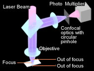
In the confocal optical system, a pinhole is provided in a position optically conjugated to a focused position (confocal plane) in order to eliminate excess light coming from non-focused positions. Only the focused point on a specimen passes through the pinhole and is detected so that clear images can be obtained without causing defocusing or flare. Olympus has adopted a circular pinhole optimized to maximize the confocal effects for higher resolution. Furthermore, a photo multiplier is used at the receiver so that very weak light can be detected, specimens with low reflectance can be observed, and the S/N ratio can be enhanced.
Scanning

With ordinary microscopes, the light coming from the condensing lens uniformly illuminates the surface of a specimen, and therefore the rays of light being reflected off and scattered from various points on a specimen overlap each other, causing flare to occur and the image quality to deteriorate. With the confocal laser microscope, a micro mirror scans minute spot light in the X and Y directions so that an image on the confocal plane only can be transmitted. This allows an image of high contrast without flare to be obtained.
Extend focus
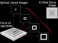
In the confocal optical system, an image is expressed as one tomogram with a shallow focal depth. Therefore, a clear image with a deep focal length can be obtained by moving the focused position up and down and forming different images in layers in the Z direction.